The fishing reel is an essential part of any fisherman’s equipment. Attached to the fishing rod, a fishing reel is how you cast and pull the fishing line. But learning how to use a fishing reel is not enough because you also need to be able to differentiate between different types of reels and know how each one is utilized.
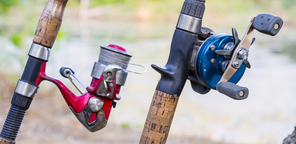
There are three basic types of fishing reels. Spincast reels are the best for beginners because of how easy they are to use. The spool is completely enclosed and you control them by pushing a special button under your thumb as you release the line. Another type is the spinning reel. Spinning reels are the most commonly used reels by anglers of all advancement levels. They are attached underneath the rod and before you cast the line you need to open the ‘bail’ – this loosens the line. You stop the line from loosening completely by holding it with your index finger and release the finger as you make the casting motion. When the bait falls underwater in the right position, you close the ‘bail’ of the reel. Spinning reels are a little bit more complicated than spincast reels but provide more accuracy. The third basic type of fishing reels is the baitcasting reel. Although notoriously difficult to control, baitcasting reels allow the angler to do long casts with utmost precision.
What makes baitcasting reels difficult
Baitcasting reels sit on top of the fishing rod and have a rotating spool. They are difficult to operate due to two main factors: the need to control the spool with your thumb as you cast and the potential for the so-called ‘backlash’. Even the best baitcasting reel will require some practice to fully learn how to operate.
First, you need to release the spool using a button or a lever to let it spin. Then, as you cast the line, you put your thumb on the spool to slow it down – that’s how you control the length and speed of the cast. The accuracy of the cast depends solely on how well you can control the spool with your thumb and this is a difficult skill to learn.
The dreaded ‘backlash’, i.e. the line getting knotted, occurs when the line is overrun or when the lure meets resistances, for example as it goes underwater. To lessen or even fully avoid backlash, you need to manually adjust the spool tension according to the size of the lure. So each time you change lure, you also need to additionally make changes in your spool settings.
If you want to catch plenty of fish you don’t necessarily need to master the craft of using a baitcasting reel. You can also use these fish finders or any other products to make fishing easier. Baitcasting reels are a great tool to learn as you gain more fishing experience but they are not essential to enjoying this wonderful sport.